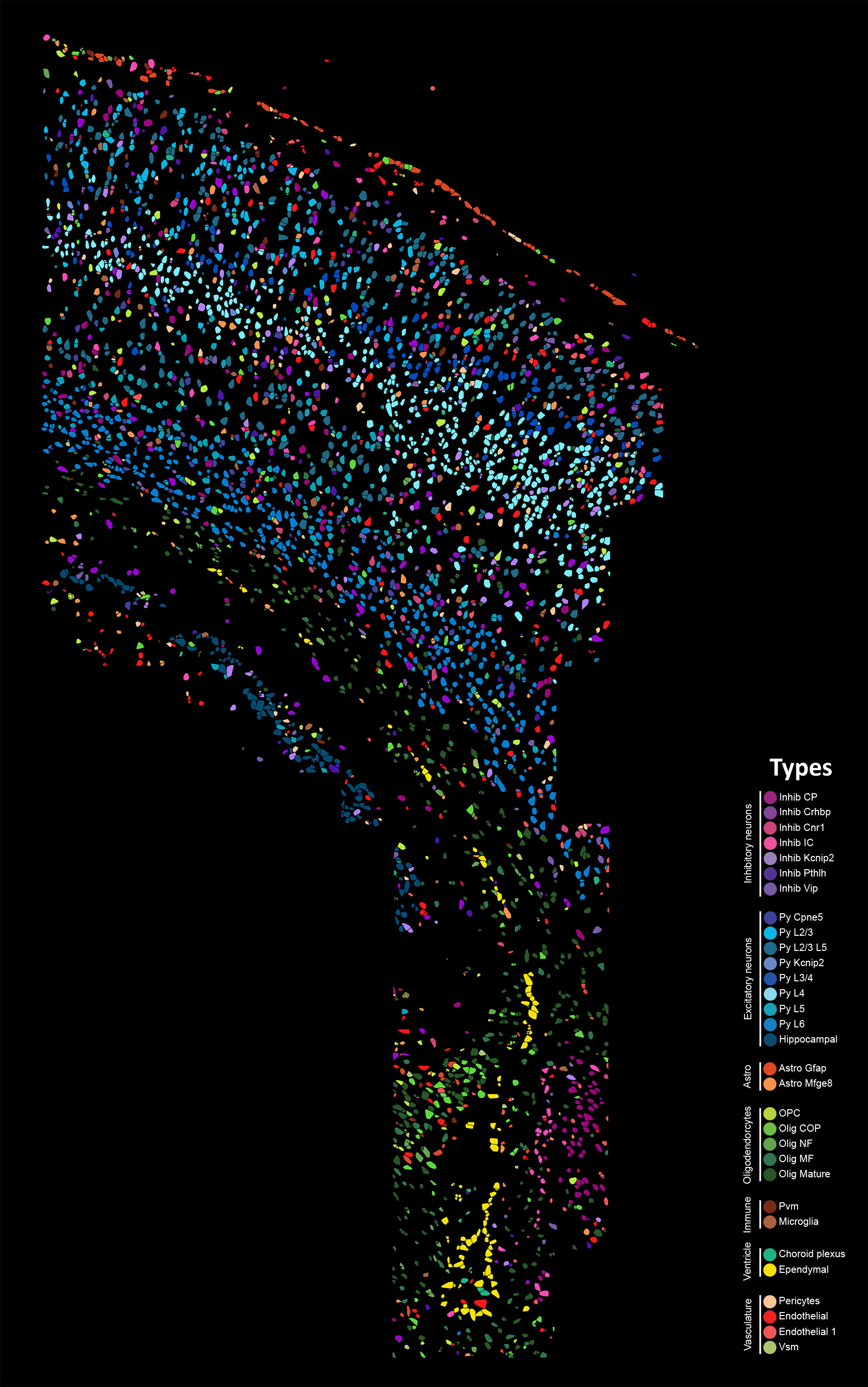
osmFISH Clusters
Cell type atlas
Major cluster and sub clusters
Below links contain images of the spatial distribution and description of the major clusters and their sub-clusters.
- Inhibitory neurons
1.1 Inhibitory CP: Spatial, Description
1.2 Inhibitory Crhbp: Spatial, Description
1.3 Inhibitory Cnr1: Spatial, Description
1.4 Inhibitory IC: Spatial, Description
1.5 Inhibitory Kcnip2: Spatial, Description
1.6 Inhibitory Pthlh: Spatial, Description
1.7 Inhibitory Vip: Spatial, Description - Excitatory neurons
2.1 Pyramidal Cpne5: Spatial, Description
2.2 Pyramidal Layer 2-3: Spatial, Description
2.3 Pyramidal Layer 2-3 Layer 5: Spatial, Description
2.4 Pyramidal Kcnip2: Spatial, Description
2.5 Pyramidal Layer 3-4: Pyramidal Layer 3-4, Description
2.6 Pyramidal Layer 4: Spatial, Description
2.7 Pyramidal Layer 5: Spatial, Description
2.8 Pyramidal Layer 6: Spatial, Description
2.9 Hippocampal neurons: Spatial, Description - Astrocytes
3.1 Astrocytes Gfap: Spatial, Description
3.2 Astrocytes Mfge8: Spatial, Description - Oligodendrocytes
4.1 Oligodendrocyte Precusor Cells (OPC): Spatial, Description
4.2 Comitted Oligodendrocyte Precusor Cells (COP): Spatial, Description
4.3 Oligodendrocyte Newly Formed (NF): Spatial, Description
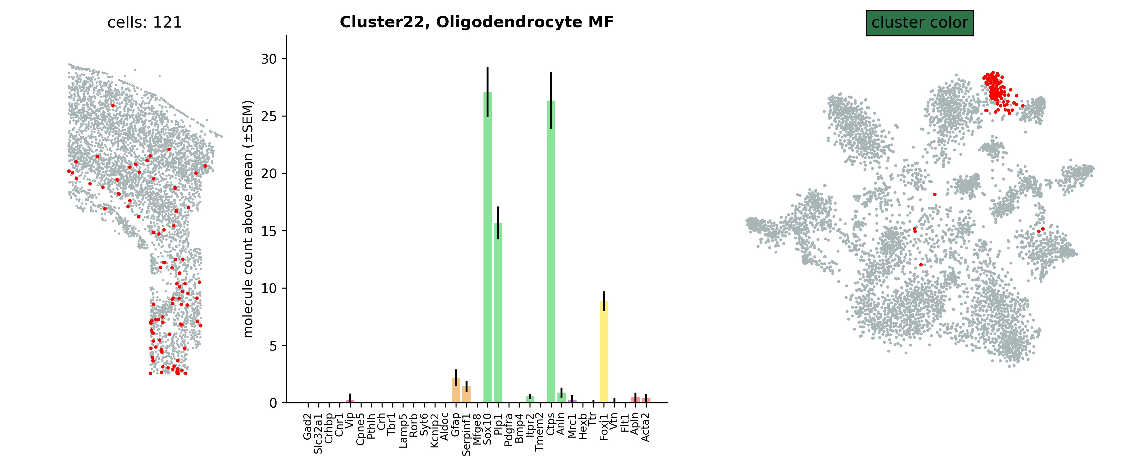
4.4 Oligodendrocyte Myelin Forming (MF): Spatial , Description
4.5 Oligodendrocyte Mature: Spatial, Description - Brain immune
5.1 Perivascular Macrophages: Spatial, Description
5.2 Microglia: Spatial, Description - Ventricle
6.1 Choroid Plexus: Spatial, Description
6.2 Ependymal: Spatial, Description - Vasculature
7.1 Pericytes: Spatial, Description
7.2 Endothelial: Spatial, Description
7.3 Endothelial 1: Spatial, Description
7.4 Vascular Smooth Muscle: Spatial, Description